SNS 358535155 - Rui Manuel de Almeida Pinheiro - Abdominal and Pelvic CT (03 June 2025)
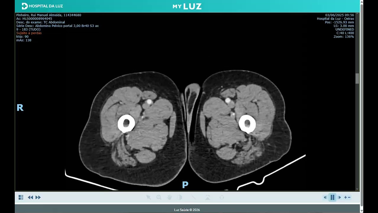
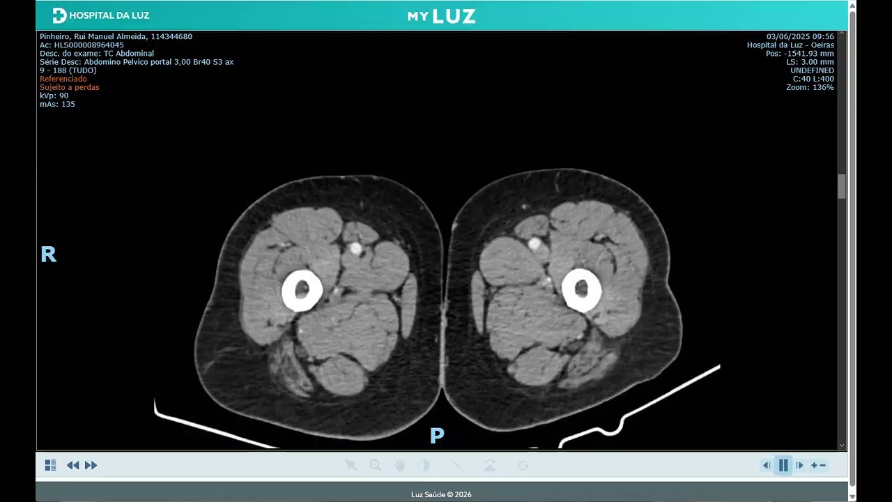
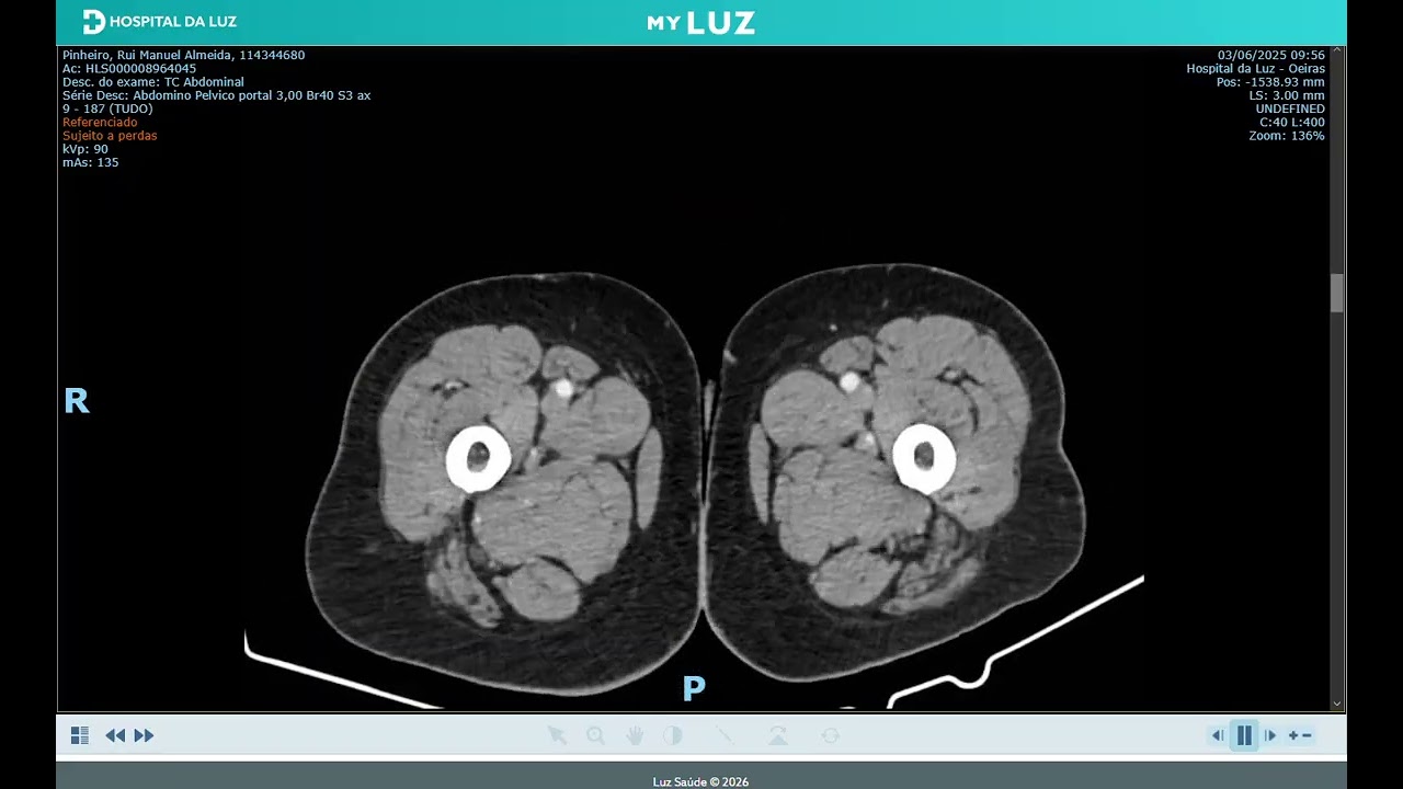
Abdominal and Pelvic Computed Tomography (CT) report dated 03 June 2025, from Hospital da Luz Oeiras, with three associated imaging videos.
CT ABDOMINAL AND PELVIC
Reason for exam
recent significant worsening of DM, intends to exclude pancreatic LOE; slight thickening of the external stem of the left SR on prior CT, intends to evaluate the SR glands.
Technical protocol
Multislice acquisition of the upper abdominal planes to the pubic planes. Oral opacification of the digestive tract. IV administration of iodinated contrast.
Reading
The liver has dimensions within normality and regular contours, with multiple small scattered hypodense mimetic images in relation to biliary cysts overlapping with those present in the study dated 24-5-2022.
Biliary ducts not dilated.
Gallbladder without signs of lithiasis with calcific density.
Pancreas, spleen and adrenals with normal CT aspects.
Small centimetric accessory spleen.
Atheromatous parietal calcifications of the aorta and iliac arteries.
Kidneys of normal dimensions, in usual topography, with globally preserved parenchymal thickness.
Bilateral renal cortical cystic alterations translated by hypodense nodular images, the most expressive on the left with 2.4 cm and on the right with 1.7 cm.
On the right we do not identify aspects of radiopaque lithiasis or dilation of the excretory system.
Small radiopaque lithic foci in the left renal sinus, non-obstructive, the most expressive with 4 mm as known.
Diverticular alterations at the level of the colon mainly sigmoid and descending in relation to known diverticulosis.
Bladder in moderate repletion, with no evidence of focal parietal or endoluminal lesions.
Globular prostate, with central calcifications.
Symmetrical seminal vesicles.
Absence of ascites or adenopathies, along the ganglion chains covered.
Degenerative osteoarticular alterations with greater expression in the vertebral column.
Note: Alterations compatible with diffuse pulmonary emphysema in the covered segments of the lung parenchyma.
Conclusion
No evidence of pancreatic or adrenal alterations detectable by this imaging technique. Pulmonary emphysema in the covered segments. Remaining aspects overlapping with those present in the prior study of 2022.
Hospital da Luz Oeiras · R. Coro Santo Amaro Oeiras, 12 · 2780-379 Oeiras · Portugal